
SIB, Lunaphore and the Geneva University Hospitals (HUG) are collaborating to enable precision cancer medicine. As part of an Innosuisse project, they aim to develop an integrative solution for the phenotypic analysis of tumors powered by automated multiplex staining, image analysis and machine learning.
“Aerial maps” of tumour samples: powerful prognosis tools
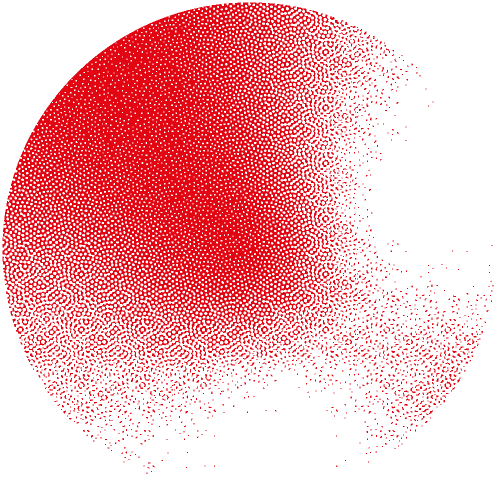
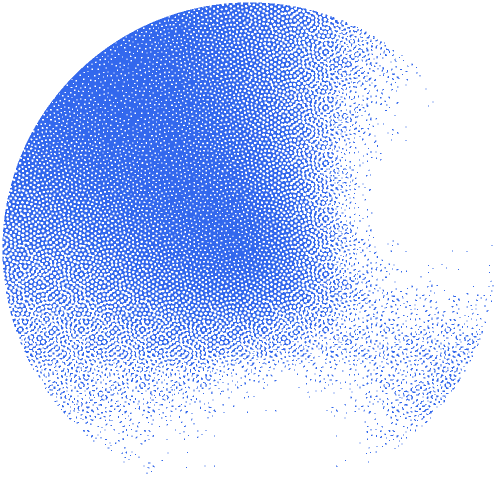
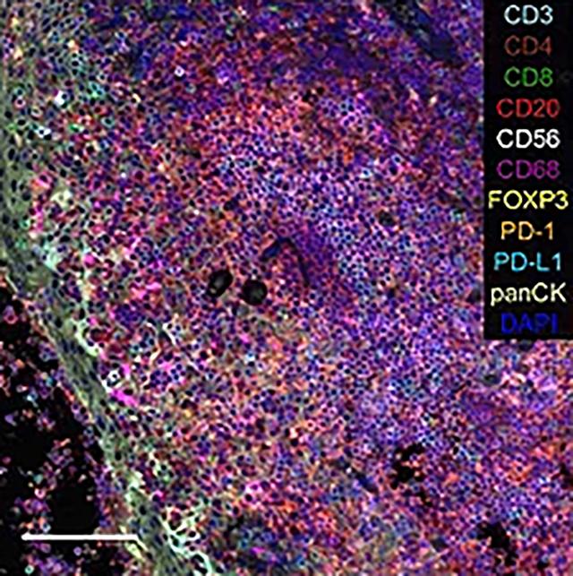
“Quantifying and localizing the various cell types and morphological features present in a tumour – a bit like aerial images of landscapes – is a key step in estimating the prognosis and the treatment of certain cancers” explains Thomas McKee, Head of the Molecular Pathology Unit at HUG. Characterizing this landscape can indeed help guide clinicians understand the prognosis and guide treatment decisions in cancer patients. “Immunostaining [see box] is the method used today to create such maps. However in most settings we are currently limited to one single stain.”
What is immunostaining?
This antibody-based method is used to detect a specific protein in a sample. In the context of this project that aims at characterizing the tumour microenvironment, it will consist of sequentially applying fluorescent markers to tumour sections, in order to identify the cell types (cancer cell, immune system cell…) present in tumours and their relations.
Integrating existing technology into a novel clinical solution
“At Lunaphore, we developed a technology enabling fast and easy immunostaining of samples allowing to visualize simultaneously more than 40 markers: this is called multiplexing”, explains Diego Dupouy, co-founder and CTO of Lunaphore.
The solution proposed in the Innosuisse project initiated in July 2020 - which involves Lunaphore, SIB and the Molecular Pathology Unit at HUG – will combine Lunaphore’s prototype, a microscope-integrated automated stainer - operated via a user-friendly software for data acquisition – together with image analysis and artificial intelligence (AI) tools to analyse the resulting images semi-automatically.
“We are excited to be bringing our machine learning, image analysis, digital pathology and software development expertise to the project”, says Aitana Lebrand, senior project manager at SIB’s Clinical Bioinformatics group and main research partner in the project, who will be working with Andrew Janowczyk (CHUV & SIB). “The solution will provide a major leap towards the analysis and generation of biomarkers for routine diagnostic usage in clinical pathology”, she concludes.